Mohammed A Hussein1*, Raghad M Mahmmed2, Afnan A Sebawaih2, Basma A El-Maghraby2, Eman E Mohammed2, Zekra S Mohamed2 and Ali A Ali3
1Biochemistry Department, Faculty of Applied Medical Sciences, October 6 University, Sixth of October City, Egypt 2Medical Labs Department, Faculty of Applied Medical Sciences, October 6 University, Sixth of October City, Egypt 3Vice president of post graduate studies, October 6 University, Sixth of October City, Egypt
*Corresponding Author: Mohammed A Hussein, Biochemistry Department, Faculty of Applied Medical Sciences, October 6 University, Sixth of October City, Egypt.
Received: April 07, 2021; Published: April 29, 2021;
Background: Resveratrol is a natural polyphenolic phytoalexin commonly found in fruits, grape and red wine. The objective of this study was to prepare RENE for Hela cell anticancer therapy using low chitosan and tripolyphosphate (TPP) method.
Methods: In the present study resveratrol-chitosan nanoparticles was prepared made in the presences of TPP using a phase inversion method. Morphology, particle size and zeta potential of RENE was then characterized. Furthermore, RENE was evaluated for its cytotoxic effect on Hela cells as well as their effect on TBARS, GSH, GST, caspase-3, NF-κB and P53.
Results: RENE was showed spherical shape with mean particle size of around 49.5 ± 0.05 nm with (+15.75) negative zeta potential. The treatment of Hela cells with RENE led to a high inhibition in the Hela cell proliferation as concluded by the low IC50 values 31.89 µg/ml. Also, RENE possess apoptotic properties via activation of the TBARS, caspase-3 and P53 protein expression as well as inhibition NF-Κb, GST and GSH levels. Conclusions: The results presented here may suggest that RENE possess anticancer and apoptotic effects on Hela cell proliferation, and therefore, can be used as new approach of pharmaceutical drugs. Also, the results clearly suggest that NF-κB, is one of the central players in the synergism of RENE and act as a regulator of caspase-3 and P53.
Keywords: Resveratrol; Resveratrol Nanoemulsion(RENE);Chitosan; Hela; NF-κB and Caspase-3
RENE: Nanoemulsion Resveratrol Nanoemulsion; TPP: Tripolyphosphate; TBARS: Thiobarbituric AcidReactive Substances; GSH: Reduced Glutathione; GST: Glutathione S Transferase; NF-κB:Nuclear Factor-κB; P53: Tumor Protein P53
Today, one of the major public health problems causing death in the world is cancer [1]. The second common cancer among women worldwide is cervical cancer [3]. Crucial genes involved in this process include APC, K-ras, DCC, p53, c-myc, COX-2, mismatch repair genes, cell adhesion genes [4,5]. Accumulation of alterations during carcinogenesis leads to impairment of normal growth inhibition by increased cell growth and by inhibition of apoptosis, resulting in clonal expansion of tumor cells [5].
Resveratrol is a common polyphenol found in many fruits; grapes and blueberries [6,7].It has been studied as cardio protective [8,9], anti-cancer [10,11], antioxidant and anti-inflammatory [12,13] effects.
The mechanism by whom resveratrol has favorable effects would be related to the ability of resveratrol to improve the bioactivity of nitric oxide and/or its inhibitory effects on COX-1 and NF-κB[15] and to block platelet aggregation with the ability of prostaglandins, synthesized with COX-1, to specifically inhibit [16] such as thromboxane A2, which is a powerful cause of platelet aggregation.
Resveratrol was used in vivo testing to determine its anti-inflammatory bowel disease [16-19]. Nevertheless, there are no reports on the effects of RENE in treatment of cervical cancer. In addition, we are reporting an easy way of preparing resveratrol-chitosan nanoparticles and assessing their apoptotic mechanism in cervical cancer cells, in as extension of our interested research program to evaluate the importance of RENE in the medicine [20-24].
Resveratrol, 5-fluorouracil, Chitosan medium molecular weight (Sigma-Aaldrich Chemical Co. catalog NO.448877-50G), Triglyceride tridecanoate (Sigma-Aaldrich Chemical Co. catalog NO.T7517-5G), Soy lecithin (Avanti Polar Lipids Catalog No. 206775), chloroform (HPLC grade), and PBS triglyceride tripalmites (Sigma-Aaldrich Chemical Co. catalogue NO.T8127-100g.). ATCC American type of culture collection obtained hela cells (human cervical carcinoma). The purchase of Sigma included dimethyl-sulfoxide, violet crystal and blue dye (St. Louis, Mo., USA). Lonza purchased Fetal Bovine serum, DMEM, RPMI-1640, L-glutamine, HEPES buffer solution, gentamycin and 0,25% trypsin EDTA. Purple Crystal Stain (1%): It was then made from 0.50 (w/v) violet crystal and 50% methanol and filtered with ddH2O by a filter paper from Whatmann No.1.
Methods Preparation of nanoparticlesThe ionotropic gelation process based on the electrostatic interaction between negative and positive charged molecules was used to create resveratrol-chitosan-TPP nanoparticles [17]. Briefly, Chitosan-TPP nanoparticles loaded with resveratrol existed in amino groups in interaction with anionic TPP salt groups. Chitosan stock solution was produced in acidified distilled water at1 mg/mL, while TPP solution was produced in distilled water at 1 mg/mL. The chitosan (1 mL) supply was first taken away and its volume was changed to 1,5 mL using distilled water for 10 minutes.
Next 5 μL tween 80 were added to the ethanol resveratrol stock (1 mg/mL). In the chitosan solution resveratrol was then added. Finally, the TPP solution as an emulsified resveratrol-chitosan solution as a cross-connector (100μL) has been added downwards. The solution obtained was removed for 30 minutes and centrifuged for 5 minutes at 4000g. The supernatant was finally moved to a new tube and kept for later analysis.
Characterization of resveratrol nanoemulsionRENE was performed by a pattern of R-25-28 o C with a D8 Advance X-ray difractometer (Bruker - Germany), with nickel (Ni) filtered with CuKα(Ţ=1.54184 A0) as an X-ray source. The resveratrol Nanoscope (RENE) had the crystalline nature and grain dimensions.
Nicolet 6700 (Thermo scientific–US) sample infrared spectrum (IR) is recorded. Thermo-gravimetric test (TGA) TGA-50 was used for thermal analysis (Shimadzu, Japan) Morphology and size of RENE were examined by Scanning Electron Microscope (SEM, JSM- 690, JEOL, Inc., Tokyo, Japan) and Field Emission Transmission Electron Microscopy (FETEM, JSM- 2100F, JEOL Inc.) at an accelerating voltage of 15Kv and 200 Kv.
High-performance liquid chromatographyRENE has been detected with a high performance liquid chromatography (Shimadzu, Tokyo, Japan) system with a C18 reverse phase column and a UV detector, with a flow rate of 1mL per minute. The Mobile Phase consisted of 500 mL of acetic acid, 2,5 mL of methanol and deionized water. The HPLC is used to quantify non-encapsulated and nano-encapsulated resveratrol concentrations. For the calculation of the peak area was used the external standardization method.
Morphology and resveratrol nanoemulsion (RENE) CharacterizationResveratrol nanomulsion morphological characteristics were examined using a microscope field Emission (FETEM, JSM-690, JEOL, Inc.) (SEM, JSM-690, JEOL, Inc.), and a 200 kV and 100.000 magnification accelerating voltage (FETEM, JSM-2100F, JEOL). A drop of nanoparticles has been covered in carbon and is permitted for 24 hours at room temperature. The TEM picture shows that the NLC and CSNLC average diameter is approximately 67 and 98 nm each and the shape is round. IR spectrum was also recorded using the KBr pellets on the Shimadzu MR 470.
Entrapment efficiencyRENE (5.0 mg), 5.0 mL distillated water and 5.0 ml of ethyl acetate, was crushed and dispersed. After the mixture was carefully mixed, the ethyl acetate phase was separated. The UV-Vis spectrophotometer has been used for the free resveratrol content of 419 nm in the phase of ethyl acetate. Equation calculated for drug entrapment efficiency(1):
Anticancer assay Cytotoxicity evaluation using viability assayThe cells were seeded in the 96-well plate at a cell concentration of 1 to 104 cells in a well in a growth medium of 100 μl for the cytotoxicity analysis. Fresh medium with various resveratrol nanoemulsion concentrations (RENE) was added after 24 hours of seeding. Serial twice dilutions of the chemical compound tested have been added to confluent, 96 well, floor-to-bottom cell monolayers dispensed in the form of a multi-chain pipette (Falcon, NJ, US).
At 37°C in an incubator moistened with 5% CO2 over 24 hours, the microtiter panels were incubated. For each test sample concentration, three wells have been used. Without test sample and DMSO, the control cells were incubated. The small amount of DMSO (maximum 0.1%) present in the wells did not affect the experiment. The viable cell yield was determined by a colorimetric method after the incubation at 37°C for 24 hours. In brief, after the end of the incubation period, media were aspirated, and the crystal violet solution (1%) was added to each well for at least 30 minutes.
The stain was removed and the plate was rinsed with tap water to remove all excess stain. Glacial acetic acid (30%) was then added to all wells and carefully mixed, and the plate absorption was then measured using a test wavelength of 490 nm after gently shaken micro-plate reader (TECAN, Inc.).
For background absorption found in wells without adding stain all results have been corrected. In the absence of the compounds tested, the treated samples were compared to the cell control. All triplicate experiments have been conducted. Each test compound was calculated for its cell cytotoxic effect. In order to determine the number of sustainable cells, the optical density was measured in the microplate reader (SunRise, TECAN, Inc.) and the viability percentage was measured as:
Viability = [(ODt/ODc)]x100%
Where ODt is the mean optical density for tested sample wells and ODc is the average untreated cell optical density. After treatment with the specified compound, the relationship between survival cells and drug concentration is drawn into the survival curve of each tumour cell line. From the graphical plots of the dose response curve for each concentrate with Graphpad Prism software, the 50% inhibitor concentration (IC50), required to cause toxic effects in 50% of intact cells (San Diego, CA. USA).
Mode of treatment1x106 cells/well of HeLa cells were sown in 96 well plates. Resveratrol nanoemulsion (RENE) (5, 10, 15, 20 and 25 μM) was treated 24-hours after incubation. The cells were scraped, washed two times with PBS, lysed for thirty minutes with 520 mL of potassium chloride (1,15%) and centrifuged at fifteen thousand minutes. The methods adapted to Cereser.,et al.[25]Ksenija.,et al.[26] and Habig and Jacoby [27] have been examined for Lipid Peroxidation, reduced glutathione, and glutathione transfers. According to the Bradford method, the concentration of cell proteins has been determined [28].
On the other hand, tests on pro-inflammatory biomarkers and caspase 3 (units/mg protein) activity were assessed using the pro-inflammatory Griess reagent at 540 nm (Protocol)[29], with the support of a platform reader from SpectraMax (Molecular Devices, CA, USA), as described in the manual of the fabricator (ELISA Company Elabscience Biotechnology, Beijing, China). The enzyme-related immuno-sorbent assay (Cayman Chemicals, Ann Arbor, MN, USA) according with the manufacturer's instruction also determined the nuclear factor kappa-B (NF-κB) p65 subunit DNA binding activity. Three times every experiment was repeated.
Western blot analysis of P53In an assay buffer on radioimmunoprecipitation for 30 min, Hela Cells (human cervical carcinoma) was prepared using the Li.,et al.method [30]. In order to remove insoluble material, lysates were centrifuged for 10 min at 15,000 x g at 4 tube. The protein was collected in the supernatant and maintained for 5 minutes at 95oC. Protein samples were transferred to polyvinylidene difluoride membranes after 10% SDS-PAGE gel electrophoresis. The membranes were incubated with anti-p53 (1:3,000) for 1 hour at 37oC after blocking 10 percent non-fat milk for 1 hour at room temperature. Three times for 5 minutes, the membranes were washed using BPS. The membranes were incubated for 2 hours at room temperature, by an incubation with an enhanced chemiluminescence detecting reagent, with rabbit (1:2,000) or goat anti-rabbit antibody (521-31-3; Pierce, Rockford, IL, USA).
Statistical analysisResults were expressed as the means ± Standard Deviation (SD). Differences between groups were evaluated by using one-way ANOVA, followed by Duncan’s test. All statistical analyses were performed using the statistical software SPSS 18.0 (SPSS Ltd., Surrey, UK).
IR spectrum infrared spectrogram of resveratrol nanoemulsionshows a phenol hydroxyl groups absorption peak at 3436cm-1 as well as benzene ring absorption peaks at 2827, 2920 exists. Light scattering techniques as well as transmission electron microscopy analysis shows that resveratrol nanoemulsion had size of around 49.5 ± 0.05 nm (Figure 1) with negative zeta potential of +15.75.
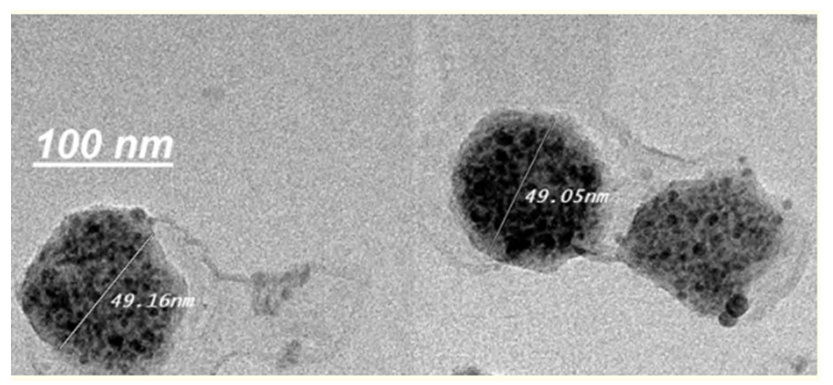
Figure 1:Morphology and size measurements of resveratrol nanoemulsion characterized by TEM are shown, with scale bar 100 nm, respectively.
As shown in the figure 2, the treatment of Hela cells with resveratrol nanoemulsion (RENE) led to a high inhibition in the cell proliferation as concluded by the low IC50 values (31.89 µg/ml) which revealed a high anti-tumor activity of the RENE against human cervical carcinoma.
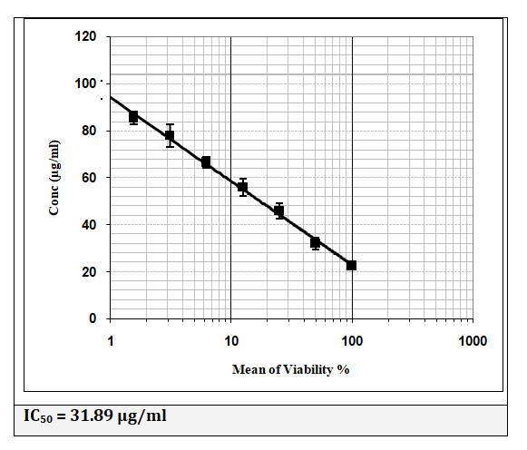
Figure 2:IC50 (%) of resveratrol nanoemulsion against Hela cells after 24 h of incubation as assayed by MTT (n = 4).
Figure 3 showed the induction effect of RENE on TBARS in Hela cells. As shown, TBARS was dramatically increased after RENA treatment at (5-25 µM). Moreover, RENA (5-25 µM) treatment also inhibit the level of GSH and activity of GST (Figure 4 and 5).

Figure 3:The effect of treatment with resveratrol nanoemulsion (RENE) (5-25 uM), on TBARS levels in Hela cells.

Figure 4:The effect of treatment with resveratrol nanoemulsion (RENE) (5-25 uM), on reduced glutathione (GSH) levels in Hela cells.
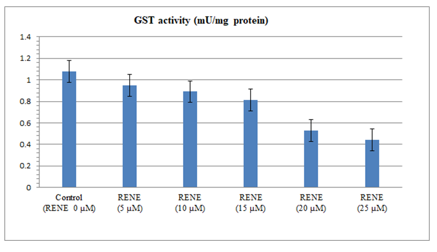
Figure 5:The effect of treatment with resveratrol nanoemulsion (RENE) (5-25 uM), on glutathione S transferase (GST) activity in Hela cells.
The results in figure 6 illustrated that RENA at (5-25 µM) produced a marked elevation of caspase activity. Taken together, these results suggested that RENE displayed rapid and potent anti-tumor effects against Hela cell lines.Also, the present study further detected the inhibitory effect of RENA at (5-25 µM) on NF-κB activity in Hela cells (Figure 7). The expression of p53 protein was increased significantly in the RENA at (5-25 µM) treated HeLa cells compared with that in the control group (Figure 8 and 9).

Figure 6:The effect of treatment with resveratrol nanoemulsion (RENE) (5-25 uM), on caspase activity in Hela cells.

Figure 7:The effect of treatment with resveratrol nanoemulsion (RENE) (5-25 uM), on Nuclear factor kappa-B (NF-κB) activity in Hela cells.

Figure 8:The effect of treatment with resveratrol nanoemulsion (RENE) (5-25 uM) on relative expression value of P53 protein in Hela cells.
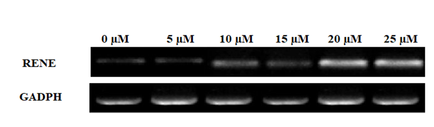
Figure 9:The effect of treatment with resveratrol nanoemulsion (RENE) (5-25 uM) on electrophoretic pattern of P53 protein in Hela cells.
Resveratrol is a promising natural compound for many diseases prevention and treatment. However, its low levels of stability and cellular bioavailability limit its anti-tumor activity. Two approaches have been used to increase its stability and bioactivities: (i) formation peracetate ester of resveratrol [31] or resveratrol -docosapentaenoic acid ester [32]; (ii) using nanocarriers such as nanoliposomes [33] and NLCs [34]. The chemical modification makes lipophilic resveratrol prodrugs, which require chemical cleavage before releasing nonencapsulated resveratrol. Nanoliposomes are not stable, and encapsulated compound can be leaked out. NLCs do not have those problems and have been widely used in pharmaceutical and nutraceutical research. There are several characteristics necessary for a chitosan to exhibit ionic interaction with resveratrol. These include:
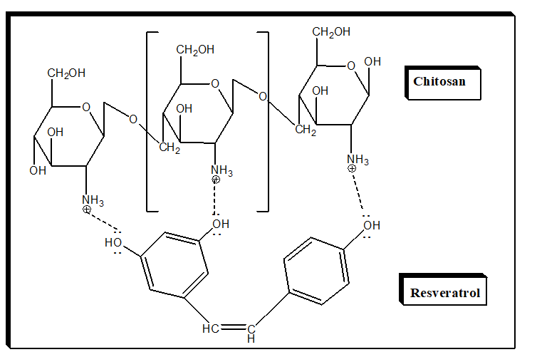
Figure 10:Proposal diagram for interaction between chitosan and resveratrol.
Infrared spectrogram of the resveratrol raw powder shows a phenol hydroxyl group absorption peak at 3252 cm-1 and benzene ring absorption peaks at 2827, 2920 exists. Infrared spectrogram of RENE shows a hydroxyl group absorption peak at 3436 cm-1 exists, but the characteristic absorption peak of resveratrol did not appear, proving that resveratrol had been completely wrapped in chitosan.
Our results in confirmed with the results of Hussein.,et al. [17]showed that the addition of chitosan in presence tripolyphosphate will lead to increase particle diameters.
To further determine the size of the RENE in the dry state, electron microscopic analysis was performed. TEM images illustrated both the size and morphology of plain NLC and CSNLC (Figure 1).
At pH 7, NH3+ of chitosan get deprotonated and changing to NH2 negatively group. Also, hydroxyl group of resveratrol which is negatively charged is no longer able to interact with NH2 group of chitosan and leading to the release of resveratrol from nanoparticles.
Anti-tumor activityUsing MTT assay, the effect of the resveratrol encapsulated (NLC) and resveratrol-chitosan coated (CSNLC) on the proliferation of Hela cells was studied after 48 h of incubation.
This study demonstrated that RENE was decrease the viability of Hela cells. From research in the field of nanotechnology, it was noted that nanoparticles improved drug solubility, controlled drug release, enhanced bioavailability, increased stability and improved long-term storage [35]. Resveratrol possesses antiproliferative effects through the induction of death in many different cell lines, including colon cancer [36]. Also, it has low solubility, stability, and bioavailability [37]. Nanoparticles were shown to improve the stability and enhancing compound’s bioavailability [38]. The results of the MTT assay with nanoparticles indicate the anticancer activity of both resveratrol formulas. This may be due to the resveratrol encapsulated nanoparticles’ rate of bioavailability. Many studies indicate that nanoparticles improve bioavailability [39]. However, if the rate of absorption is too great, it may lead to toxicity on cancer cells. This could be the issue in the current study.
Cells withstand and counteract oxidative stress using several and different defense mechanisms, ranging from free radical scavengers and antioxidant molecules/enzymes to sophisticated and elaborate DNA repair mechanisms to reduce excessive levels of ROS and prevent irreversible cellular damage [40,41]. An increased rate of ROS production occurs in highly proliferative cancer cells, owing to the presence of oncogenic mutations that promote aberrant metabolism. Increased oxidative stress is well documented in transformed cells [42-45]and growing evidence suggests that ROS act as second messengers in intracellular signaling cascades which induce and maintain the oncogenic phenotype of cancer cells [46]. During cervical carcinogenesis an increase of oxidative DNA damage, as shown by progressive increase in the levels of 8-OHdG from normal to SIL to invasive carcinomas, has been reported [47]. In our recent work we demonstrated that RENE-induced oxidative stress and decrease the GSH level and GST activity which could be provoked an increased and selective protein oxidation in keratinocyte cells transfected with the whole HPV-16 genome [48].
Apoptosis can be started through two distinct pathways, the death receptor (extrinsic pathway) mitochondrial mediated (intrinsic pathway). Both lead to activation of the caspase cascades "executioner caspases" such as caspase-3, -6 and -7. The active executioner caspases bind cellular substrates, leading to the characteristic biochemical and morphological changes. This is followed by chromatin condensation, nuclear shrinkage and DNA fragmentation. Binding of cytoskeletal proteins leads to cell fragmentation and formation of apoptotic bodies [17,49,50]. As shown in the present results caspase-3 were significantly increased in Hela cells treated with the RENE compared to untreated cells.Many research studies have demonstrated that resveratrol can induce cancer cell death in cellular studies [51-53]. This is a promising attribute in the field of cancer research. It is important for the area of nanoparticles to perfect precise targeting of cancer cells, and optimal time release.
In regard of resveratrol antitumoral properties, several studies were conducted on certain cancer cell lines [17]. Cervical cancer cell lines were often used as a preferred carcinogenic model to understand molecular targets and mechanism of resveratrol action. Here are reported all the in vitroadvances for the use of RENE in the treatment of cervical cancer. In the present study we explain the effect of RENE on NF-κB activity and P53 protein expression in cervical cancer cells. we demonstrated that inhibition of NF-κB activity and activation of P53 protein expression induced by RENE leading to increase cell-death. According with this study Bava.,et al. [54] showed that other polyphenolic compounds sensitizes cervical cancer cells to the therapeutic effect of taxol, acting in the down-regulation of both NF-κB and serine/threonine kinase AKT pathway, a survival signal related to NF-κB.
Prevention of DNA oxidation is also achieved by these polyphenols mainly by quenching free radicals and modulating bio-metabolism enzymes [13]. This study demonstrated that RENE was more potent anticancer agent against Hela cell line due to the nanoparticles improved drug solubility, controlled drug release, enhanced bioavailability, increased stability and improved long-term storage [17].This study, indicate that NF-κB, is one of the central players in the synergism of RENE and act as a regulator of caspase-3 and, P53 instead is the regulator of NF-κB, which through the phosphorylation of MAPKs regulates a set of survival signals.
In the present study, we used the chitosan to prepare resveratrol nanoemulsion. Also, characterization of obtained spherical nanoparticles was evaluated using different procedures. RENE showed significant anticancer activity against Hela cells line. Also, RENE possess significant apoptotic properties via inhibition of GSH, GST and NF-κBas well as activation of the level of TBARS, caspase-3 activity and P53 protein expression. Overall, our results suggested that RENE could be a promising new approach for anticancer therapies.
Ethical approval for the data collection was granted by the Research Ethics Committee at the Faculty of Applied Medical Sciences, October 6 University, Egypt (No. 20201206).No lab. animals or Humans were used studies that are base of this research, in-vitro experiments only were used.
The authors gave consent for their data to be used in the article.
Supporting data will be made available as it contains spectroscopic and analytical data to prove the structure of obtained nanoparticles as well as their in vitro antitumor and antiapoptotic activities.
The authors declare no conflict of interest, financial or otherwise.
The authors are not currently in receipt of any research funding.
Citation: Mohammed A Hussein., et al. “NF-κB, Caspase-3 and p53 Pathways are Involved in Resveratrol-Chitosan Nanoparticles-Induced Apoptosis in Hela Cells with Oxidative Stress". Acta Scientific Nutritional Health 5.5 (2021): 128-137.
Copyright: © 2021 Mohammed A Hussein., et al. This is an open-access article distributed under the terms of the Creative Commons Attribution License, which permits unrestricted use, distribution, and reproduction in any medium, provided the original author and source are credited.


 Open Access by
Acta Scientific is licensed under a Creative Commons Attribution 4.0 International License
Open Access by
Acta Scientific is licensed under a Creative Commons Attribution 4.0 International License
Based on a work at https://actascientific.com
ff
