Mousumi Das1, Urmita Chakraborty1, Moonmoon Sinha1, Anirban Kool1, Suraia Parveen1 and Satadal Das2*
1Virology Lab, Dr. Anjali Chatterjee Regional Research Institute, CCRH, Kolkata, India
2Department of Microbiology and Molecular Biology, Peerless Hospital and B. K. Roy Research Centre, Kolkata, India
*Corresponding Author: Satadal Das, Professor, Department of Microbiology and Molecular Biology, Peerless Hospital and B. K. Roy Research Centre, Kolkata, India.
Received: January 05, 2021;; Published: February 17, 2021
Citation: Satadal Das., et al. “Newer Conventional and Nonconventional Treatment Strategy of Chikungunya Fever along with a Review of its Virology and Epidemiology”. Acta Scientific Pharmaceutical Sciences 5.3 (2021): 13-23.
Chikungunya fever, a re-emerging viral disease affecting both old and new world countries is caused by Chikungunya virus, an RNA virus belonging to Alphaviridae. Although mortality is less but the suffering from the disease is too much, even it may continue for several months. Thus, a drastic treatment protocol should be followed to contain the suffering from the disease. However, there is no specific antiviral drug, and supportive treatment strategy in acute, post-acute, and chronic phases are quite different. It is also special in pregnancy and new born. Even with all these elaborate plans, the treatment outcome is often dismal and human suffering continues. There is some treatment prospect of this disease in alternative medicine, which can be combined with the treatment plan of conventional medicine without any deleterious effect. Thus, this integrated approach may help to restrain the enormous suffering of the patients from the disease, at least with greater relief to the patient. Along with the treatment part a review of the biology and epidemiology of the virus is also included.
Keywords: Chikungunya Fever; Conventional Treatment; Alternative Medicine; Chikungunya Virus Epidemiology
Chikungunya fever (CHIKF) is a viral disease transmitted by mainly two species of mosquitoes - Aedes aegypti and A. albopictus. Chikungunya Virus (CHIKV) was first detected in 1952, during a febrile outbreak on the Makonde Plateau, a border area between Mozambique and Tanzania [1]. CHIKV usually spreads easily in populated areas with no herd immunity. Although most of the cases of CHIKF cures spontaneously, but serious complications and even death may also occur.
It is difficult to separate clinical presentations of CHIKF from other arthropod-transmitted alphaviruses like Barmah Forest virus, Ross River virus, Semliki Forest virus, O’nyong-nyong, Mayaro, Sindbis and similar viruses. The name chikungunya means “that which bends up” or “to become controlled”- due to bend down appearance following arthritis, which occurs in this disease [2]. Several million of CHIKF cases have already been reported mainly from Asia and Africa, as well as from Europe, the islands of Indian Ocean and the Americas.
CHIKV is a small (60 - 70 nm) spherical enveloped virus with single- stranded positive sense RNA molecule and belongs to the genus Alphavirus, which included more than 30 species of arthropod borne viruses [1], and the family Togaviridae (Figure 1). There are three structural (C, E1, E2) and four non structural proteins in CHIKV [2]. Alpha viruses are not found in the Antarctica, and they are also sensitive to temperature above 58°C. Ecological pattern and vector transmission of Alpha viruses is similar to many Flaviviruses like dengue fever virus and yellow fever virus.
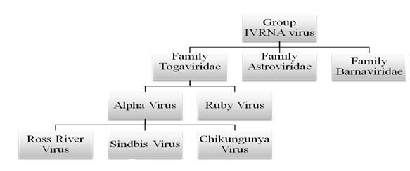
Figure 1: Diagram shows the RNA viruses/Togaviridae family viruses/Alphaviruses [Modified from 41].
In 1823 illness similar to Chikungunya was described from Zanzibar, in local Swahili language it was known as kidinga pepo, which means a cramp-like spasm. After one year an epidemic of this disease was observed by Christie, which was published later in the British Medical Journal. Following this in successive years Chikungunya-like disease was observed in St. Thomas Island, New Orleans and part of South Carolina of the United States in 1827 to 1828 [7]. About 1000 CHIKF cases were found in European and American travelers who returned from these areas at that time [8].
CHIKV is now included in the re-emerging virus group, particularly due to its recent expansion in the Americas. Generalized neuro-developmental delay was recorded in 51% of CHIKV infected children in La Reunion while only 15% of the control cases showed similar changes [3]. Outbreaks of CHIKV in other areas highlighted its capability of undergoing mutation, resulting increased infectivity, and spread with variable clinical presentations in areas without any herd immunity [4]. Sporadic as well as epidemic CHIKF is rampant in Senegal to Cameroun as well as in Nigeria, Uganda, Malawi, Burundi, Democratic Republic of Congo, Angola, Guinea, Central African Republic and South Africa. Many epidemics occurred in the 1960s and then in the 1990s in different Asian countries like Thailand, Vietnam, Burma, Indonesia, India, Sri Lanka, Timor and the Philippines [5]. In the year 2004, a massive outbreak of CHIKV occurred in Kenya, where seroprevalence rate reached 75% (Lamu Island), and then it spread to the Comores, Seychelles, and Mauritius islands. Many immigrants from the Comores carried the disease which spread to some countries in the Indian Ocean, including the La Réunion island where epidemic occurred in March-April of 2005 [6]. After this, Local CHIKV transmission in the Americas was found in December 2013.
Travelers act as carriers which helped spread of the disease between countries. Thus, this group of carriers may act as sentinel population, from where we can study the biological behavior of the pathogen and the disease kinetics of such a pathogen [11]. If we consider the Americas, the disease kinetics of CHIKV remains unknown. The current trend indicates transmission of human to mosquito. Still, it is not clear whether an enzootic cycle is present in susceptible non-human primates in this area, in which CHIKV viremia occurs. There is no important manifestation of the disease in non-human primates, which is usually found with other viruses like yellow fever virus infection. Studies are required to evaluate the actual role of non-human primates, but also the other potential reservoirs like rodents, birds, or other vertebrates need to be identified to maintain CHIKV in the environment [12,13].
World Health Organization (WHO) 2008 guidelines on CHIKF estimated that, complete resolution occurred in 87.9%, episodic stiffness and pain occurred in 3.7%, persistent stiffness without pain occurred in 2.9%, and persistent painful restriction of joint symptoms occurred in 5.9% cases. Time required for complete recovery from CHIKF varies with age, in younger patients it is 5 - 15 days; in middle aged patients it is 1 to 2.5 months, and in the elderly patients it is longer [14]. The residual Musculo-skeletal complications cause hardship to the bourgeoning population of India, especially because this disease is more prevalent in socioeconomically weaker section, who are not living in hygienic surroundings, and cannot afford costly medical treatment for this disease. In Delhi CHIKF epidemic occurred in October 2010. The number of reported cases increased from 33 on October 19, 2010 to 1000 on 1st November 2010 [15].
CHIKV genome and structureCHIKV is an enveloped positive-sense single stranded RNA virus, with icosahedral symmetry. It is a member of the family Togaviridae (Figure 1) and it belongs to the genus Alphavirus. O’Nyong-nyong, Mayaro and Ross River viruses, which also can cause diseases in human beings and they have similar antigenic pattern [16]. The length of genome of CHIKV is about 12 kb which contains two open reading frames (ORFs) - 5´ORF and 3´ORF. The 5´ORF is translated from genomic RNA responsible for non-structural (ns) proteins P1-P4 and sub-genomic RNA derived the 3´ORF, responsible for structural proteins envelope (E1 and E2), capsid (C), and two peptides (E3 and 6K) (Figure 2) [17]. The diameter of the cubicle viral particle is about 70 nm with 240 capsid proteins, surrounded by an lipid envelope. The envelope contains 80 spikes of E1 and E2 glycoprotein [18].
Alphaviruses enter to the target cells by endocytosis and few receptors like DC-SIGN, L-SIGN, heparin sulphate, laminin and integrants implicated in this process, but their precise roles have not been clearly proven. The endosome is acidic causing structural changes in the envelope, which leads to exposure of E1 peptide after endocytosis followed by fusion of virus and host cell membrane. This helps cytoplasmic delivery of the core and release of viral genome [19]. Further two non-structural proteins are translated from the viral mRNA, and cleavage of these to generate nsP1 to nsP4. These proteins initiates viral replication synthesizing an intermediate negative-strand RNA, sub-genomic (26S) and genomic (49S) RNAs [20]. In the sub-genomic RNA synthesis, the expression of the C-pE2-6K-E1polyprotein precursor, which is processed by auto-proteolysis, after releasing of capsid, the pE2 and E1 glycoproteins generates by further processing. Pre E2 and E1 together move to plasma membrane from the Golgi body, and in plasma membrane pE2 is cleaved to E2 and E3. Then the nucleocapsid is attached to viral RNA and envelope glycoproteins are engaged. The alpha-virus particle assembled with an icosahedral core, and buds at the cell membrane [19,21]. The 60 - 70 nm size viral particles are enclosed in envelopes, containing genome with 11805 nucleotides (Figure 2). Initially the non-structural proteins are expressed and sub-genomic RNA is generated. This leads to a new step of transcription of structural viral proteins, and replicates of the viral genome on the RNA template.

Figure 2: General structure of chikungunya virus as well as genomic configuration and proteins expressed [Modified from 49].
Comprehensive epidemiologyArboviruses are transmitted through the bite of haematophagous arthropods to humans, and other animals. Disease is produced in human beings and animals by five main Arbovirus families - Bunyaviridae, Togaviridae, Flaviviridae, Reoviridae and Rhabdoviridae [22]. Some species of arbovirus like Dengue virus, West Nile virus, yellow fever virus, Mayaro virus, Saint Louis encephalitis virus, Venezuelan equine encephalitis virus, Oropouche virus and Rocio virus are well known to cause human diseases in Brazil [23]. In 1952 an epidemic of CHIKF occurred in Tanzania, and the disease was also prevalent in many countries of Asia, Africa, Europe and the Americas [24]. In the epidemic of this disease in 2004, many African countries were affected [25]. In 2004 the disease began in the Indian Ocean islands, Seychelles, Mauritius, Comoros, Mayotte, and La Reunion [26], where from 2005 - 2007, 2,66,000 cases affecting 34% of the island population was reported [27]. In 2006, CHIKF was reported in many countries of Europe like France, Germany, Italy, Switzerland, Belgium, and England [28]. In Americans, first endemic CHIKV infection was reported in Saint Martin Island, in 2013 [29]. Later in 2015, some cases were found in Brazil, Colombia, Bolivia, Paraguay, Ecuador, Venezuela, and many other countries. Pandemic strain with Asian genotype of CHIKV was found in Brazil in September 2014. At the same month, another outbreak caused by a different genotype (ECSA), occurred in Feira de Santana. In 2014, 3,657 cases of CHIKF had been reported in Brazil. Two genotypes of the virus is now prevalent in Brazil with 94% population is at risk. At present, three viruses cause epidemics in Brazil - dengue, CHIK and Zika viruses. High temperature, humidity, rainfall help vector proliferation and assist in transmission of these viruses. Climate change in tropical countries along with migration of people, diminished forest area, unplanned urban areas and poor sanitary conditions are important factors which lead to viral spread and transmission.
Mosquitoes of the genus Aedes mainly Aedes aegypti usually transmit the disease very efficiently. This effectiveness is principally on the grounds that this class is exceptionally anthropophilic and lives in closeness to people. A. albopictus is well known as the second mosquito which can also transmit the disease. A change related with an amino acid replacement in the glycoprotein E1-A226V permitted the infection more readily to adjust in the vector, in this manner expanding its capacity to spread the infection. This finding was seen in the strain of CHIKV that circulated during an episode in the Indian Ocean islands, alluded to as the Indian Ocean genealogy [31]. Different types of mosquitoes, from various parts of the world, can transmit CHIKV, including Culex annulirostris, Anopheles stephensi, Mansonia uniformis, Eretmapodites chrysogaster, and Opifex fuscus [30]. Transmission through these vectors is identified with their topographical appropriation and the kinds of transmission cycles, regardless of whether wild or metropolitan. Notwithstanding vector transmission, the vertical transmission of CHIKV has been distinguished. On Reunion Island, 7,509 pregnant ladies were observed for such transmission. In view of polymerase chain reaction as well as IgM antibody results, 678 of them were found infected. Of these, 39 showed viraemia during intrapartum period and roughly 49% of their infants were found infected [32]. Haematogenous transmission of these viruses in the viraemic period may cause a significant issue for donating blood in endemic territories. In La Reunion epidemic, 47 units of donor blood out of 37,750 were found positive for CHIKV [33]. Similarly, when CHIKV RNA and IgG antibody was studied in Puerto Rico in 2014 epidemic of the disease, 2.1% of the donor blood units were found positive while about 25% donors were infected and sero-converted [34].
The outbreaks were reported in Madagascar from December 2006 - July 2009. After a couple of years in the 2009 and 2010 in La Réunion again the disease reappeared followed by re-importation to Europe [9]. Again in between 2011 - 2013, about 11,000 cases were found in the Republic of Congo, 29 cases were reported from Rajasthan, India, 1500 cases were found in Cambodia, 633 cases in Papua New Guinea, and 1100 cases in Philippines during the period [10]. In between 2011 - 2013 a gigantic flare-up with an excess of 11,000 cases occurred in the Republic of Congo (Brazzaville). Present geographical distribution of the disease is shown in figure 3.
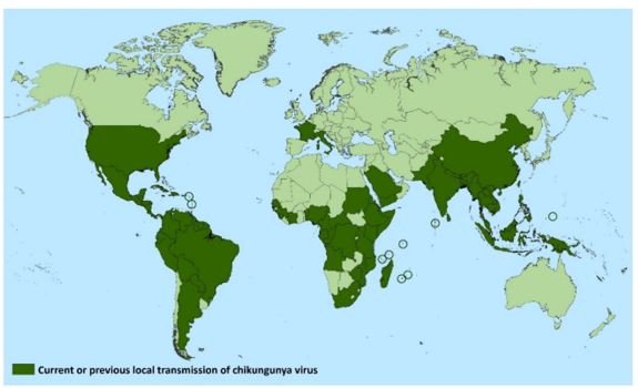
Figure 3: Recent geographic distribution of CHIKV [49].
Clinical spectrum of CHIKV infectionFollowing the bite of the infected mosquito the virus first enters the skin, replicate in fibroblasts, and then enters the blood and disseminated to the liver, spleen, muscles, lymph nodes, brain and joints. Within the first week of infection the viral load may become as high as 109 copies/mL in blood, however, viremic period stay for 2 - 10 days. Like other viral diseases, both asymptomatic and symptomatic cases are found in this disease which are presenting mild clinical spectrum to a severe form of the disease with disabling conditions.
Asymptomatic infection: The strain variation and capacity of sero-conversion is reflected to the number of asymptomatic infections in various age groups. The asymptomatic infection can vary from one epidemic to another. In Mayotte islands, the Comoros and in India sero-conversion in asymptomatic cases were 28, 14 and 17.5%, respectively. 12-month community-based prospective study revealed 82% asymptomatic infections developed in the Philippines [31]. In Managua, Nicaragua, 58.3% asymptomatic infections reported in another community-based study in children 2 - 14 years of age.
Symptomatic infection: Symptomatic cases are usually categorized in three phases - acute, post-acute, and chronic.
Acute phase: In this phase the first three weeks of the disease is considered after an incubation period of 2 - 4 days. The infected individuals express high fever, polyarthralgia/polyarthritis, intense myalgia, and it is often accompanied by headache, photophobia, rash etc. Mild Polyarthralgia to severe polyarthritis even with disability are typical manifestation of CHIKV infection. Mainly the polyarthralgia affects limb joints symmetrically and bilaterally, and they become typically swollen. During the acute phase it is the most frequently observed clinical manifestation in adults. Stiffness in the distal joints, particularly the interphalangeal joints of the hands and feet, wrists and ankles upon awakening are usually present [35]. If myalgia is also present particularly in the forearms, arms, calves and thigh, it may compromise physical activities to a great extent. Skin affection is observed with variable frequency, they may be expressing in up to 80% of cases, and it involves primarily the face, trunk, and extremities. It often follows 2 - 5 days of arthralgia and myalgia and persists for 2 - 3 days. Skin manifestations are mainly itchy or non-itchy macular and maculopapular erythematous rashes with facial oedema; however, in acute phase, vesicles, bullae, increased skin pigment, photosensitivity, exfoliative dermatitis, erythema nodosum are also seen. Preexisting psoriasis and oral ulcers may show increased activities [35,36]. Clinical presentations of cervical lymphadenopathy, nausea, vomiting, diarrhea, chills and weakness may be found. The acute phase commonly involve central nervous, urinary and respiratory systems of our body with exacerbations of pre-existing chronic autoimmune, respiratory and cardiovascular diseases [37].
Post-acute phase: In this phase the clinical manifestations begin after the 21st day and it continues for three months [38]. The clinical course of the disease is paradoxical - initial transient improvement is usually followed by polyarthralgia or polyarthritis for a long period and the affected patients have to continue pain reducing medicines for a prolonged period [38]. The prolonged polyarthralgia following the acute phase is dependent on genetic susceptibility, treatment modality, age, sex etc [38]. In most published papers it has been observed that in about 50 - 90% patients it may continue even after second or third week of the disease depending on age (> 40 years), sex (> in females), degree of pyrexia (> high pyrexia), number of affected joints (> if six or more joints show arthritis), viremia and depression [38].
Post-acute phase complications like arthralgia, arthritis (synovitis with or without effusion), bursitis, periostitis, tendinitis and tenosynovitis etc. proceed continuously or intermittently with precipitating factor like cold. Decompensation of pre-existing osteoarthritis or tendinitis may occur. Among other complications local oedema; carpal, tarsal, cubital tunnel syndromes; morning joint stiffness, neuralgic pain, vascular hypersensitivity like Raynaud syndrome etc. may also occur. Non-specific clinical manifestations like chronic fatigue, skin color alteration, fall of hairs, resurfacing of pre-existing disease manifestations of metabolic, endocrine, cardio-vascular and psychiatric diseases are also commonly found.
Chronic phase: If arthralgia continues for more than 3 months the disease is diagnosed as a chronic phase disease. In Chikungunya 40 to 80% patients can progress to the chronic phase, and they may undergo clinical manifestations for a few months or even several years [38]. In this phase there are wide variations of clinical manifestations, arthritis may be migratory, and however, typical signs of inflammation are usually absent. An important chronic phase study up to 10th month was done in India with 200 patients. It was found that joint pain, fatigue, and neuritis persisted in 46%, 13%, and 6% patients respectedly, and in MRI study of the joints of some patients changes were noted. The patients in the chronic stage have been classified into three groups as per disease progression: i. the major group, cured with/without treatment, ii. patients with persistence of the disease for long time, iii. patients with serious condition [38]. Tenosynovitis is the most common of musculoskeletal manifestations observed during chronic phase of the disease in some patients. The most affected parts are wrist, finger joints, extensors and flexors involving two or more tendons. A majority of the patients with hypertrophic wrist tenosynovitis encounter nocturnal paraesthesia in fingers. After the acute phase hypertrophic tenosynovitis with some bilateral syndromes are usually observed including cubital, carpal and tarsal tunnels. The quality of life is highly affected after CHIKV infection. Mortality has been noticed frequently among the CHIKV infected patients with co-existing diseases [39].
In the epidemic of La Reunion Island in 2005 - 2006, there were 232 deaths [26] and 58 deaths were recorded in Colombia in 2014-15 [29]. The better understanding regarding patient assessment has changed with mortality rate among the acute phase patients and its association with symptoms and infection.
Laboratory diagnosisThe common laboratory diagnosis of the infection include direct isolation of the virus, molecular biology based tests such as RT-PCR, serological tests by the detection of specific IgG/IgM antibody by ELISA etc [27] and relates to the phase of the disease (Figure 4). For isolation of virus directly, samples are usually collected within eight days after onset of sign and symptoms. Various cell lines are commonly used for in vitro culture including Vero, BHK-21 and HeLa etc. and the cytopathic effect are observed after three days post infection. Although isolation of viral particle is confirmatory, it is time consuming and requires extra set up. Alternatively, immunofluorescence or RT-PCR is performed accordingly for confirmation. RT-PCR techniques have limitation as it only detect viral RNA. However, RT-PCR is considered more important for early diagnosis for newborns with meningo-encephalitis and vesiculobullous der¬matitis [41]. Among the serological tests available point-of-care (POC) immunochromatographic tests and ELISA detects IgM within fifth day after onset of symptom and IgG antibody after few days [27]. These rapid tests are advantageous over other molecular tests as these do not require laboratory equipments even refrigerator.
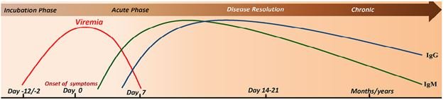
Figure 4: Sequence of events during CHIKV infection. Infected Aedes mosquito deposits the virus into the dermis and epidermis of the skin. The viral infection is characterized by an incubation period which is followed by the acute phase during which a rapid rise in viremia occurs, and clinical symptoms such as joint pain, fever, maculopapular and petechial rash appear. As the viral load increases the host innate and adaptive immune responses are evoked simultaneously. IgM and IgG levels rise and the virus is resolved from the host. The virus may persist in synovial fluid even after the induction of strong immune response. This fact leads to the altered joint pathology and progression of the chronic phase for months to years [modified from 47].
TreatmentIn the treatment schedule prepared by a multi-professional group [42] for pain in acute phase, visual analogue scale (VAS), with scoring from 0 - 10 is used, where 0 and 10 indicates two extreme indicators of pain. However, it should be interpreted in association with clinical examination particularly for stress as indicated by the French work group [46]. During history taking of the patients, history of allergy and drug reactions should be noted along with presence of any co-morbidity like diabetes, hypertension, renal diseases, glaucoma, cardiomyopathy etc. Steroids and non-steroidal anti-inflammatory drugs (NSAIDs) should be avoided except in associated neuropathy or encephalopathy. Absolute bed rest and proper hydration should be maintained. Any adverse reaction due to the medicines should be closely observed [43].
When VAS score is less (1 - 3), paracetamol (500 - 750 mg 4 times a day) and dipyrone (1.0g 4 times a day if body weight is more than 60 kg) may be used [43]. When VAS score is moderate (4 - 6) dipyrone and paracetamol may be used together. Tramadol can be used if there is allergy to dipyrone. Care should also be taken in pregnancy and breast feeding conditions. In negative response to treatment the pain may be assessed again with DN4 questionnaire again [43].
When there is severe neuralgic pain 25 mg amitriptyline may be added with other drugs. One may also try gabapentin (300 mg twice a day) or pregabalin (75 mg twice a day). Anticonvulsants and antidepressants should be used carefully in these patients. If there is history of cardiac arrhythmia, amitriptyline should not be used. When VAS score is high (7 - 10), tramadol or codeine (30 mg 4 times a day) may be combined with other drugs, while supportive treatments for nausea and constipation may also be given [43]. Again, if there is a negative response, the DN4 questionnaire should be used after 7 days of treatment and if this indicates absence of neuralgic pain corticosteroids or NSAIDs may be used considering co-morbid disease if any. In pregnancy NSAIDs should not be used. Different studies indicated ambiguity regarding presence of the virus in the newborns [38].
Newborns and children: When the mother is suffering from the disease the newborns should be observed for at least 5 days in NICU [38]. Children with the disease are similarly treated as adults except NSAIDs and Codeine.
Treatment of pain in the post-acute phase: This phase extends from 22 - 90 days with variable clinical manifestations. The main aim of treatment in this period is to relieve pain and neuropathy which may be assessed by DN4 questionnaire, and if required anti-depressants or anticonvulsants may be used along with psychological support. During the post acute or mild-intensity pain phase (VAS from 1 to 3), NSAIDS are well known therapeutics for relieving pain such as ibuprofen (400 mg) every 8h, nimesulide (100 mg) or meloxicam (7.5 - 15 mg) or naproxen (500 - 750 mg) may be used every day for 7 - 10 days. If pain persists steroids like prednisone (up to 0.5 mg/kg body weight) each day may be used and should be continued for 3 - 5 days more followed by tailing of 5 mg weekly [43].
Chronic phase treatmentThe persistence of clinical manifestations for more than three months is considered a chronic phase. In some patients intense inflammatory manifestations similar to rheumatoid arthritis are observed which according to the VAS are categorized under mild to moderate intensity pain therapeutic recommendations, as mentioned earlier. In case when the pain intensity is very strong other drugs such as disease modifying anti-rheumatic drugs (DMARDS) like methotrexate and hydroxychloroquine may be used [42]. However, other additional approaches of relieving pain and inflammation can also be encouraged to reduce joint pains. In acute phase, rest is mainly recommended as physical activities may aggravate inflammation.
NSAID precautionsBefore NSAID treatment careful measures regarding any existing co-morbidities should be investigated. Evaluation of creatinine, transaminase and urea levels, in both pre and post usage of NSAIDS should be done. Care should be taken with associated comorbidities like cardiac diseases, diabetes, hepatic diseases, hypertension, peptic ulcer, renal diseases etc. In case of gastric injuries gastric protector may be used. During moderate and severe pain (VAS score 4 - 10), corticosteroids are the best known therapeutics such as Prednisone. However, NSAIDS may also be used with an opioid such as tramadol following the above mentioned doses. Prednisone has been known to exhibit recovery from pain of patients suffering with arthralgia. For local inflammatory lesions which do not respond properly to oral medication such as bursitis, capsulitis, synovitis etc. administration of topical anti-inflammatory or corticosteroid injection to the affected region is recommended. Moreover, during synovitis it is recommended to avoid surgical decompression for relieving recurrent pain due to poor healing ability and algodystrophy [38].
In chronic phase if there are evidences of intense inflammatory reaction, according to the American College of Rheumatology it may be similar to rheumatoid arthritis. Methotrexate and hydroxychloroquine may be considered in this condition with caution [42]. Sometimes some local measures like NSAID gel patch; release of joint fluid or steroid injection, physiotherapy may help to relieve pain [38]. Some antiviral agents like ribavirin, arbidol, interferon, chloroquine, furin inhibitors, and favipiravir are found effective in vitro, however, as such still there is no specific antiviral drug for this disease.
Although some vaccines are prepared with inactivated virus, but they showed very low immunogenicity and are also associated with adverse reactions. Vaccines with chimeric viruses, electroporated DNA, recombinant antigens, viral-like particle, attenuated virus by large-scale codon etc. are not standardized. Again, the doctors are not familiar to the disease and laboratory diagnosis facilities are only available in reference centers. A chart showing pharmacological management of arthralgia in chronic CHIKF has also been given elsewhere [48].
Effect of homeopathic medicines on CHIKVConventional treatment is symptomatic and it usually includes anti-inflammatory drugs such as ibuprofen and naproxen, paracetamol (acetaminophen), rest and fluids. A comparative evaluation of treatment of CHIKV in different systems of medicine (Allopathy, Ayurveda, Homeopathy and Traditional) was observed by Dilip., et al. and concluded that, “...all the systems of medicine are equally important for the management of Chikungunya”, indicating an integrated approach. A study on CHIKV with homeopathic treatment in 532 patients was conducted in Kerala by Biju and Sarathchandranon, indicating a good outcome where nine homeopathic medicines were used (Ledum palustre, Ruta graveolens, Rhus toxicodendron, Belladonna, Eupatorium perfoliatum, Bryonia alba alba, Apis mellifica, Formica rufa and Arsenicum album). Rao and Rejikumar., et al. also carried out a study of homeopathic prophylaxis in CHIKV [44-46].
Homeopathic treatment of CHIKF is one of the most utilized alternative therapies. The selection of medicines is based upon the individualization and symptoms similarity of every individual. Homeopathic medicines are aimed to treat Chikungunya, addressing its root cause and individual susceptibility.
The homeopathic medicines for CHIKF are selected depending on the intensity of the symptoms which should be done by a qualified homeopathic physician. There are many medicines in homeopathy which can be used in CHIKF. These include: Eupatorium perfoliatum, Pyroginum, Rhus tox, Cedron, Influenzinum, China, Arnica, Belladonna, Bryonia, Polyporus pinicola etc. Eupatorium perfoliatum is a well known preventive medicine for Chikungunya and the 200C potency is commonly used. Eupatorium Perfoliatum mother tincture is also known to be effective in debilitating joint pains, however, different potencies may be used as per the intensity of the symptoms. Arsenic album (higher potency) and China may be used in severe condition. In severe headache, China, Belladonna, Cedron may be used. China is indicated in headache and prostration.
Rhus toxicodendron: Rhus tox homoeopathic preparations can mediate with histamine, prostaglandins and other inflammatory mediators. It can prevent Chikungunya fever arthritis (CFA) induced in rats along with body weight and haematological changes. Rhus tox gives protection from radiological joint changes in CHIKF. Anti-arthritic with decreased arthritic pain scores were observed with Rhus tox. In a study on 1061 CHIKF cases by Rejikumar., et al. in Kerala, the Eupatorium perfoliatum 200C gave protection in 82.19% cases [50,52].
Polyporus pinicola: Polyporus pinicola diminished joint pains and headache in CHIKF. Congestion in head with hot flushed face; constant nausea, with severe pain in phalangeal joints, wrists, ankles, knees particularly in night time, along with restlessness and intense sweating are indicated for the use of this medicine. Presence of back pain and pain in shin bone are also benefitted from the use of this medicine.
Bryonia: This homeopathic medicine is mainly indicated in CHIKF where joint pain increases with motion and in severe joint pain when the joints are hot and swollen. Increased thirst and dry mouth if present in the suffering patient then it is specifically indicated. In a paper published in Indian Journal of Research in Homoeopathy (IJRH), Bryonia alba 30C was found to prevent CHIKF - in 2525 persons out of 19750 Bryonia alba 30C treated cases CHIKF occurred, while 2919 out of 18479 placebo treated cases manifested the disease indicating a 19.76% relative risk reduction. Bryonia alba 30C was also used as genus epidemicus in Kerala where decreased incidence of CHIKF occurred; however, this needs to be verified in similar epidemics [51].
Eupatorium perfoliatum: It is indicated in CHIKF where severe pain is present in bones. Eupatorium perfoliatum specially indicated when patient suffering from CHIKF feels that his or her bones are broken and become restless and cry and in cases where there is chill. After taking the medicine there is relief of pain followed by perspiration. This medicine is also indicated if there is vomiting after chill. A conceptual map of homoeopathic treatment of arthralgia in chikungunya [48] has also been described.
Belladonna: In children suffering from CHIKF Belladonna is indicated. It is specifically indicated when there is fever without thirst, feet are cold, head sweating, joint swelling, intense pain in arm and leg, erythema or pallor of skin, trembling gait, light induced headache, insomnia and drowsiness are present.
Arsenicum album: CHIKF cases with restlessness and irritability, when the patient is scared, aggravated in midnight and after exposure to cold rainy weather or cold food, this medicine is indicated. CHIKF with twitching of the limbs, coldness, vomiting, sweating, fainting, oedema of feet etc. are also specially indicated to treat with this medicine.
Asafoetida: It decreases pain of the joints after recovery from CHIKF. It is also indicated when there is severe bone pain, night time throbbing pain in different parts, when symptoms are increased in warm weather in morning time, and better feeling with cold materials.
Pyrogenium: As an antiseptic homeopathic medicine, it reduces fever, restlessness, chill, vomiting, and weakness in morning time.
Due to extensive suffering of the Chikungunya fever patients a combined integrative treatment approach of conventional and alternative medicine may be beneficial to the patients suffering from this disease. This integrative approach should be planned by Government authorities of the countries where the disease is endemic.
Copyright: © 2021 Satadal Das., et al. This is an open-access article distributed under the terms of the Creative Commons Attribution License, which permits unrestricted use, distribution, and reproduction in any medium, provided the original author and source are credited.


 Open Access by
Acta Scientific is licensed under a Creative Commons Attribution 4.0 International License
Open Access by
Acta Scientific is licensed under a Creative Commons Attribution 4.0 International License
Based on a work at https://actascientific.com
ff