Vinicius Cidral Correa1*, Renan Akira Miyashiro1, Lucas Moraes Nunes Martins1, Cinthya Laysa Gehrke1, Bernardo Kaplan Moscovici1,2 and Nelson Chamma Capelanes1,2
1Unidade Paulista de Oftalmologia - UPO, São Paulo, SP, Brazil
2Departamento de Oftalmologia e Ciências Visuais, Universidade Federal de São Paulo - Unifesp, São Paulo, SP, Brazil
*Corresponding Author: Vinicius Cidral Correa, Unidade Paulista de Oftalmologia - UPO, São Paulo, SP, Brazil.
Received: July 19, 2021; Published: July 30, 2021
Corneal ulcer represents an ophthalmological problem of great importance given its possible causes, therapeutic challenges and repercussions. As a definition, a corneal ulcer is a solution for discontinuity of the Korean epithelium associated with variable involvement of the underlying stroma. Corynebacterium sp represents an optional, non-mobile, commensal gram-positive, aerobic or anaerobic bacterium. In this way, its pathogenic power is linked to predisposing corneal factors and the virulence of the pathogen to result in corneal damage. This case report a male patient, 74 years old, diagnosed corneal lesion during the COVID- 19 pandemic season.
Because of the above, it is concluded that there is a need for early intervention with the use of adequate propaedeutic with the identification of the causative agent, despite the prolonged empirical treatment, associated with a follow-up with a maximum of 48 hours between visits.
Keywords: Cornea; Ulcer; Corynebacterium sp
Corneal ulcer represents an ophthalmological problem of great importance given its possible causes, therapeutic challenges and repercussions. Since the cornea alone corresponds to the greater refractive power of the human eye; the visual deficit involved results in significant comorbidity for the patient.
As a definition, a corneal ulcer is a solution for discontinuity of the Korean epithelium associated with variable involvement of the underlying stroma. It represents the second cause of unilateral blindness, being surpassed only by cataracts in developing countries [1].
In our national reality, studies show that the incidence of corneal ulcers is higher in males, between 30 to 39 years, with ocular trauma as a major risk factor followed by the inadequate conservation and maintenance of contact lenses [2]. Bacteria are the most isolated microorganisms in corneal ulcers, especially Gram-positive ones. Among bacterial causes, 90% are caused by 4 main groups: Micrococcaceae, Streptococcus sp, Pseudomonas sp and members of the Enterobacter family [3]. Among the bacteria in the conjunctive microbiota, the main ones are coagulase-negative Staphylococcus and Corynebacterium sp, which have low pathogenicity. This characteristic leads, in general, to infecting corneal ulcers resulting from chronic eye diseases or eye injuries.
The incidence of Corynebacterium sp in corneal ulcers is variable. However, a study by M. Srinivasan and collaborators shows a 12.5% representation [3]. The same study highlights Corynebacterium Xerosis as the main representative of the genus.
Corynebacterium sp represents an optional, non-mobile, commensal gram-positive, aerobic or anaerobic bacterium. In this way, its pathogenic power is linked to predisposing corneal factors and the virulence of the pathogen to result in corneal damage. Corneal defense factors are: the intact corneal epithelium; immunoglobulins, free radicals and enzymes such as tear film lysozyme; beta-lysine and lactoferrin and the power to clean the eyelids when blinking [3]. However, when failing, they act as facilitators of corneal ulcer. Also, Corynebacterium diphtheria can penetrate the intact epithelium.
Corynebacterium has gained importance in recent years due to studies, such as the Prasad Eye Institute, reporting an increase in the number of eye infections due to Corynebacterium [4].
A male patient, 74 years old, diagnosed with controlled diabetes mellitus using insulin; with a history of retinal detachment and surgery to correct the right eye resulting in blindness. Assisted during the COVID-19 pandemic season.
He entered the ophthalmology service on 03/31/2020, with a history of ocular hyperemia in his right eye for 15 days. The patient postponed the search for ophthalmological care due to the prescription of exposure and contagion by COVID-19 since at the time of care the city of São Paulo was at peak incidence of cases. Since, the patient has clarification of his increased risk being of risk group, being elderly and diabetic. Visual acuity in the right eye without luminous perception. She reported using eye drops of 1% prednisolone acetate and 0.5% moxifloxacin for 3/3 hours in the 48 hours before the appointment; associated with doxycycline 100 mg 12/12 hours orally together with the therapeutic contact lens, on suspicion of neurotrophic keratitis due to diabetes mellitus. Initial biomicroscopy showed corneal lesion 4 x 6 mm, stained with fluorescein, 3 x 3 mm, whitish, dense stromal infiltrate, reaching deep stroma, thinning in the region around 50% temporal and 30% central and conjunctival hyperemia 2 + / 4 +. Referred for culture collection, remaining 12 hours without using eye drops before, keeping only Doxycycline. Soon after, a fortified antibiotic was started (Vancomycin 50 mg/ml and Ceftazidime 50 mg/ml).
Follow-up 48 hours later, the condition worsened, with corneal edema, central thinning and intense collagenolysis. Then, under a slit lamp, cyanoacrylate glue was applied to the thinning site. Reassessed in 24 hours, showing formed anterior chamber, improvement of pain, hyperemia, corneal edema and infiltrate.
A new evaluation in 72 hours, reported that the contact lens came out. Biomicroscopy: 3 x 4 mm de-epithelization, with 2 x 2.5 mm stromal infiltrate, opaque cornea, without hypopyon, without infiltration in the anterior chamber, presence of central descemetocele and absence of glue. Given it, a new application of Cyanoacrylate was performed and 12/12 hour atropine eye drops were started for pain control.
Follow-up every 48 hours, maintained with a progressive decrease in depth of the lesion and ocular hyperemia, with the glue remaining in place. Fortified and doxycycline suspended after a week, and 0.5% prophylactic gatifloxacin started 12/12. It used a thousandth corticoid 0.1% fluorometholone acetate 8/8 hours for 14 days.
On 04/18/2020, corneal scraping culture was released, with the result showing microorganism Corynebacterium sp.
Still in follow-up at the service with the use of therapeutic contact lens and 0.5% Gatifloxacin for 12/12 hours prophylactic, maintaining an anterior chamber formed with glue in a thinning area.
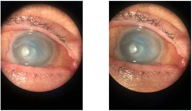
Figure
Worldwide, infectious keratitis is a cause of great morbidity and low visual acuity due to corneal opacity, ocular perforation and endophthalmitis [1-3,5,6]. The correct diagnosis of the etiologic agent is of great importance for choosing the specific treatment for each case.
Thus, the analysis of the epidemiology and periodicity of cases of corneal ulcer helps the ophthalmologist to define which agent is most likely and to choose the initial therapy when the microbiological analysis is not available at the time of service or while waiting for the result of the culture. Taking into account the clinical case, we note that the etiologic agent was Corynebacterium sp, which meets the literature.
The bacteria most often involved in a corneal infectious process are Streptococcus pneumoniae, Coagulase-negative Staphylococcus and Staphylococcus aureus. Gram negative bacteria, mainly Pseudomonas aeruginosa, and Klebsiella pneumoniae are also reported to cause corneal infection. The eye affected by the bacterial ulcer had a history of retinal detachment with the need for vitrectomy, leaving it with no luminous perception.
Thus, the biggest concern was the possibility of ocular perforation, since corneal opacity would not affect the patient's vision. Then, cyanoacrylate glue was applied due to corneal thinning. Cyanoacrylate, when in contact with water, quickly polymerizes and solidifies, forming a plate that supports the healing and epithelization of the underlying tissue, inhibits inflammatory cell migration, delaying tissue necrosis and has bacteriostatic action [6]. In some cases, even if combating infection is being effective (as in our case), collagenolysis, due to the inflammatory process, can generate intense necrosis and ocular perforation.
In our case, cyanoacrylate glue prevented this process and the patient continued with good recovery. Due to the postponement of the ophthalmic consultation due to the COVID-19 pandemic, it caused a delay in the recovery of the corneal ulcer, so questions were raised, how many patients will have worsening of their chronic ocular pathologies due to the COVID-19 pandemic? [7-10].
Because of the above, it is concluded that there is a need for early intervention with the use of adequate propaedeutic with the identification of the causative agent, despite the prolonged empirical treatment, associated with a follow-up with a maximum of 48 hours between visits.
It is also perceived the importance of cyanoacrylate glue to prevent coronal puncture, in addition to the bacteriostatic effect, helping to resolve ulcer infection. In the case seen, visual preservation was not sought, since the patient was already without luminous perception due to previous diseases.
Citation: Vinicius Cidral Correa., et al. “Cornea Ulcer Caused by Corynebacterium Sp-Case Report".Acta Scientific Ophthalmology 4.8 (2021): 33-36.
Copyright: © 2021 Vinicius Cidral Correa., et al. This is an open-access article distributed under the terms of the Creative Commons Attribution License, which permits unrestricted use, distribution, and reproduction in any medium, provided the original author and source are credited.


 Open Access by
Acta Scientific is licensed under a Creative Commons Attribution 4.0 International License
Open Access by
Acta Scientific is licensed under a Creative Commons Attribution 4.0 International License
Based on a work at https://actascientific.com
ff
