AS Zotov, AS Balalin*, SV Balalin, AM Marukhnenko, TG Efremova, SM Purshak and IA Melikhova
S. Fyodorov Eye Microsurgery Federal State Institution, Volgograd Branch, Russia
*Corresponding Author: AS Balalin, S. Fyodorov Eye Microsurgery Federal State Institution, Volgograd Branch, Russia.
Received: June 11, 2021; Published: June 23, 2021
Purpose: To assess the role of microperimetry in follow-up and treatment of patients with macular holes. Materials and Methods: Retrospective study of the minimally invasive vitrectomy results using 25G or 27G technologies in 59 patients (59 eyes) with idiopathic macular holes (IMH) was performed. The examination included the determination of the best corrected visual acuity (BCVA), tonometry, perimetry, ultrasound biometry, optical coherence tomography, fundus photography, microperimetry.
Results. After surgical treatment all patients showed a significant improvement in BCVA and retinal photosensitivity (p < 0.05). A formula was derived for the dependence of BCVA after treatment on the initial retinal photosensitivity and the maximal IMH size, which can be applied to predict the results of surgical treatment.
Conclusion: Microperimetry is a modern non-invasive examination method that allows with a higher density and resolution to localize central defects of the visual field and to carry out thorough monitoring before and after surgical treatment. The study of the retinal photosensitivity in the macular region and the maximal IMH size before treatment allow to predict BCVA in the postoperative period.
Keywords: Idiopathic Macular Holes; Microperimetry; Minimally Invasive Vitrectomy; Optical Coherence Tomography
Idiopathic macular hole (IMH) is an acquired disease that leads to decreased central vision, metamorphopsias and central scotoma. The prevalence of IMH is approximately 3.3 per 10,000 population, but the rate increases 10 times in people over the age of 65 [1].
J.D. Gass classification of macular holes was used in the study [1]:
According to the theory developed by J.D. Gass, the leading role in the pathogenesis of IMH is attributed to vitreoretinal traction.
The surgical treatment is the only treatment method for patients with IMH, in particular minimally invasive three-port vitrectomy 25G and 27G with membrane peeling, aimed at correcting the anatomical defect, which in the long term determines the improvement of visual functions [2]. According to numerous studies, microperimetry is the most reliable diagnostic method for determining the functional parameters of the retina. It makes possible to estimate the threshold of the retinal photosensitivity in a specific area of the macula or paramacular zones with the subsequent transfer of these data to the fundus image [3-5]. Microperimetry is based on laser scanning ophthalmoscope technology and retinal tracking, which allow to observe the retina in real time during the functional examination and project a certain light stimulus to a selected point [6-13].
To assess the role of microperimetry in follow-up and treatment of patients with macular holes.
Retrospective study of 25-27G vitrectomy results in 59 patients (59 eyes) aged 52 to 80 years with idiopathic macular holes (IMH) was carried out. The clinical group included 24 women and 35 men.
Inclusion criteria:All patients underwent extended ophthalmologic examination before and after treatment, including determination of BCVA (Phoropter 16625B, Reichert/Leica, USA), tonometry (NT-530, NIDEK CO. LTD., Japan), perimetry ("Perigraf Perikom", SPETSMEDPRIBOR, Russia), ultrasound biometry (А/В AVISO, Quantel Medical Aviso, France), optical coherence tomography (RS-3000 Advance2/RS-3000 Lite2, NIDEK CO. LTD., Japan), photo-registration of eye fundus (CX-1, Canon Inc., USA), fundus microperimetry (MAIA, CenterVue, Italy).
Values of retinal photosensitivity according to fundus microperimetry on MAIA microperimeter: normal values: 25 dB to 36 dB; borderline conditions: 22 dB to 24 dB; pathology: 21 dB and below.
Fixation stability is determined on microperimeter by fixation stability index, which is based on the following parameters: if more than 75% of the fixation points are within a circle of 2 degrees diameter located in the "center of gravity" of all fixation points, the fixation is classified as stable; if less than 75% of the fixation points are within a circle of 2 degrees diameter, but more than 75% of the fixation points are within a circle of 4 degrees diameter, the fixation is classified as relatively unstable; if less than 75% of the fixation points are within a circle of 4 degrees diameter, the fixation is classified as unstable. P1 and P2 according to microperimetry data represent the percentage of fixation points located within circles with diameters of 2 and 4 degrees.
All patients underwent minimally invasive vitrectomy using 25G or 27G techniques.
Statistical calculations were performed using STATISTICA 10 (StatSoft, USA) and Numbers (Apple Inc., USA) for quantitative attributes: BCVA, photosensitivity of the macular area (dB), stability of fixation (%), maximal macular hole diameter (μm). According to the small access, the nonparametric Wilcoxon criterion was used to determine differences between the pre-op and post-op results. Differences were considered statistically significant at p < 0.05.
The patients were divided into 4 groups according to the Gass classification in order to assess the treatment results. Lamellar macular hole was diagnosed in 15 patients (15 eyes), stage 2 full-thickness macular hole in 12 patients (12 eyes), stage 3 - in 15 patients (15 eyes), stage 4 - in 17 patients (17 eyes).
The average values of the pre-op results are presented in table 1. The table shows that as the IMH stage increases, there is a significant decrease in BCVA and retinal photosensitivity according to microperimetry data.
|
Indicators Groups |
Number of eyes |
BCVA pre-op |
Microperimetry pre-op |
||
|
Photosensitivity of the macula, dB |
P1, % |
P2, % |
|||
|
Lamellar macular hole |
15 |
0,36 ± 0,08 |
23 ± 0,8 |
78,8 ± 11,5 |
97,2 ± 1,6 |
|
2nd stage of IMH |
12 |
0,24 ± 0,08 |
21,6 ± 1,2 |
83 ± 14 |
94 ± 6 |
|
3rd stage of IMH |
15 |
0,18 ± 0,08 |
22,2 ± 1 |
71,9 ± 6,1 |
92,1 ± 3,7 |
|
4th stage of IMH |
17 |
0,14 ± 0,06 |
18,6 ± 1 |
59 ± 12 |
91 ± 5,4 |
Table 1: Mean values of the visometry and microperimetry pre-op results, М ± σ.
Retinal photosensitivity pre-op depended on the IMH size (Figure 1): the larger the maximal IMH diameter, the lower the patients' retinal photosensitivity was.
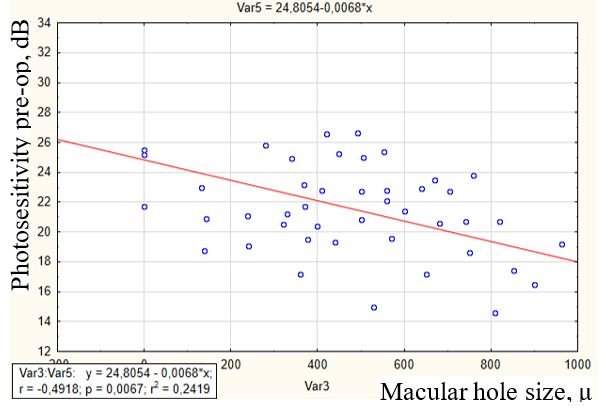
Figure 1: Dependence of retinal photosensitivity on the macular hole size pre-op.
After minimally invasive vitrectomy, complete closure of the macular hole was achieved in all cases. There were no intra- and postoperative complications.
Table 2 shows the mean BCVA and photosensitivity values after minimally invasive vitrectomy. After surgical treatment there was a significant improvement of BCVA and retinal photosensitivity.
|
Indicators Groups |
Number of eyes |
BCVA pre-op |
Microperimetry pre-op |
||
|
Photosensitivity of the macula, dB |
P1, % |
P2, % |
|||
|
Lamellar macular hole |
15 |
0,42 ± 0,09 (+17%) |
24,2 ± 1 (+5%) |
86,2 ± 5,1 (+9%) |
97,4 ± 1,6 (+0,2%) |
|
2nd stage of IMH |
12 |
0,38 ± 0,09 (+58%) |
22,6 ± 1,1 (+5%) |
95,5 ± 4,5 (+13,1%) |
99,5 ± 1 (+5,5%) |
|
3rd stage of IMH |
15 |
0,3 ± 0,09 (+67%) |
22,9 ± 0,9 (+3%) |
73,8 ± 7,1 (+2,6%) |
92,4 ± 2,3 (+0,3%) |
|
4th stage of IMH |
17 |
0,28 ± 0,12 (+100%) |
21,4 ± 1,4 (+15%) |
68 ± 11,8 (+13,2%) |
92,5 ± 4,7 (+1,6%) |
Table 2: Mean values of visometry and microperimetry post-op results, М ± σ.
In the 1st group, patients significantly improved BCVA by 0.06 (p < 0.01) and retinal photosensitivity by 1.2 dB (p < 0.05). P1 increased by 9% (p < 0.01), P2 increased by 0.2% (p < 0.05).
In the 2nd group, patients' BCVA improved by 0.14 (p < 0.01) and retinal photosensitivity by 0.06 dB (p < 0.05). P1 increased by 13.1% (p < 0.01), P2 increased by 5.5% (p < 0.05).
In the 3rd group, the patients had significantly improved BCVA by 0.12 (p < 0.01) and retinal photosensitivity by 0.09 dB (p < 0.05). P1 increased by 2.6% (p < 0.01), P2 increased by 0.3% (p < 0.05).
In the 4th group, patients had a 0.14 (p < 0.01) improvement in BCVA and a 2.8 dB improvement in retinal photosensitivity (p < 0.05). P1 increased by 13.2% (p < 0.01) and P2 increased by 1.6% (p < 0.05).
Considering the improvement of BCVA in all clinical groups after minimally invasive vitrectomy, it was supposed, that was a possibility of post-op BCVA predicting basing on data of the pre-op retinal photosensitivity and maximal macular hole diameter. Figure 2 shows the dependence of postoperative BCVA on the pre-op microperimetry indicators and macular hole size.
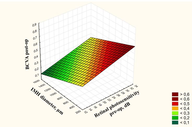
Figure 2: Dependence of post-op BCVA on the pre-op microperimetry values and macular hole size.
Based on a multifactor correlation analysis, the dependence of post-op BCVA on the pre-op photosensitivity and the maximal macular hole size was revealed, described with following formula:
Z = 0,2787 + 0,011 × X – 0,0002 × Y, where
Z - BCVA post-op, X - value of the retinal photosensitivity in the macular area pre-op (dB), Y - maximal macular hole diameter pre-op (μm).
The formula shows that higher pre-op retinal photosensitivity value and smaller the IMH diameter are associated with higher the post-op BCVA can be predicted. This formula may be used to predict the results of surgical treatment.
Further research on the prediction of IMH treatment results will be continued using data from optical coherence tomography with an angiography program.
Microperimetry is a modern noninvasive method of examination, which allows to localize central visual field defects with higher density and resolution, to carry out careful monitoring before and after surgical treatment.
The study of retinal photosensitivity in the macular area and the maximal macular hole size pre-op allows to predict BCVA in the postoperative period, which has a practical importance.
Citation: AS Balalin., et al. “Role of Microperimetry in Observation and Treatment in Patients with Macular Holes".Acta Scientific Ophthalmology 4.7 (2021): 44-48.
Copyright: © 2021 AS Balalin., et al. This is an open-access article distributed under the terms of the Creative Commons Attribution License, which permits unrestricted use, distribution, and reproduction in any medium, provided the original author and source are credited.


 Open Access by
Acta Scientific is licensed under a Creative Commons Attribution 4.0 International License
Open Access by
Acta Scientific is licensed under a Creative Commons Attribution 4.0 International License
Based on a work at https://actascientific.com
ff
