A Manton*, W Saldana and F Peck
Eastbourne District General Hospital, England, United Kingdom
*Corresponding Author: A Manton, Eastbourne District General Hospital, England, United Kingdom.
Received: May 24, 2021; Published: June 23, 2021
A male 86 year old pseudophakic patient with a poorly controlled end stage primary open angle glaucoma presented to the emergency department with an unprovoked, spontaneous expulsive suprachoroidal hemorrhage. An evisceration was conducted under general anaesthetic with a good surgical outcome.
Keywords: Glaucoma; Spontaneous Suprachoroidal Hemorrhage; Bacterial Keratitis; Evisceration
Expulsive suprachoroidal haemorrhage is a devastating condition that may be secondary to incisional surgical procedure or spontaneous. The spontaneous form is rare, however several cases have been described in the literature [1-6]. It normally occurs in the context of glaucoma and corneal disease [7]. Other risk factors include advancing age, uncontrolled hypertension, vascular disease, anticoagulation, presence of infection and chronic topical steroid use [8].
Due to the devastating nature of this condition visual recovery is thought to be unlikely and evisceration or enucleation of the affected eye is indicated. In this report, we describe a case of spontaneous expulsive suprachoroidal haemorrhage (SESH), the methods of treatment and their follow-up results.
A 86 year old male end stage primary open angle glaucoma patient presented to the emergency department with an history of severe pain, loss of vision and bleeding from the left eye. There was no recent history of ocular trauma. Previously the patient had been managed for primary open angle glaucoma with bimatoprost once at night and timolol/dorzolamide combination twice daily. I-lube and hyloforte were three times daily each to treat evaporative dry eye. One month previous, visual acuity was hand movements. Central corneal thickness was 497µm with a pressure of 30mmHg Goldman applanation tomography. Coexisting pseudophakia, myopia and untreated meibomian gland dysfunction were noted within the eye. In 2013, phacoemulsification and intracapsular lens implant were conducted without complication. A histology-confirmed basal cell carcinoma was removed from the medial canthus with Hugh’s flap reconstruction in 2018 with resulting entropion and trichiasis. Further to this, multiple bacterial keratitis episodes were managed, the most recent being in 2019 requiring admission with intensive antibiotic drops with a pseudomonas keratitis. Past medical history of hypertension, atrial fibrillation and diabetes is recorded with management of Ramipril, Atenolol, Aspirin, Insulin.
The previous evening he reported eye pain sometime after administering his usual eye drops. The following morning he awoke with transient eye pain with noticeable bleeding from the eye. At time of review there was an absence of ocular pain. On examination, visual acuity was no perception of light in the affected eye and 1/60 pin hole in the contralateral eye. Intraocular contents were prolapsed through a corneal rupture. A spontaneous expulsive haemorrhage was diagnosed (See figure 1). The eye was eviscerated, left without implant and achieved a good surgical outcome.
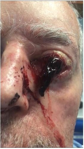
Figure 1: A colour photograph showing the spontaneous suprachoroidal haemorrhage with prolapse of intraocular contents and almost complete loss of corneal tissue.
Suprachoroidal haemorrhage is a recognised complication of incisional surgery. Here blood haemorrhages from the long or short ciliary arteries into the potential space between choroid and sclera [9]. Two putative mechanisms are suggested for this complication following decompression. These are direct rupture of the ciliary arteries due to changing ocular shape or secondary to change in shape due to suprachoroidal effusion [10,11]. Either of these mechanisms are possible if a corneal injury such as an undiagnosed bacterial corneal perforation decompresses the eye, particularly given the raised intraocular pressure. Two further mechanisms have been suggested specifically for spontaneous suprachoroidal haemorrhage. These are rupture of ciliary arteries due to inflammatory necrosis [12]. Finally, as a result of primary haemorrhage of the ciliary arteries secondary to vascular disease [13]. Contributing factors to the severity of this injury are underlying systemic hypertension, use of aspirin and probable arteriosclerosis secondary to diabetes [8].
As this was an unobserved injury the aetiology is contentious. Previous studies have highlighted risk factors such as raised intraocular pressure due to chronic glaucoma and unobserved topical steroid use [14]. It is also important to note that this cornea was previously injured both through multiple bacterial keratitis episodes, including a recent pseudomonas keratitis which is known to be able to penetrate through an intact epithelial layer [15] and through clear corneal incisions at time of phacoemulsification. Either of these could provide structural weakness and shearing planes once a suprachoroidal haemorrhage occurred. While most cases of SESH in the literature describe patients with both corneal disease and glaucoma [16], Srikanth., et al. report a case with neither predisposing risk factor [4]. It is postulated that glaucoma contributes to the risk of SESH by three mechanisms. 1) via bullous keratopathy that may become infected and lead to perforation, 2) relatively greater decompressive force at perforation and 3) weakening of the arterial wall caused by focal ischaemia of ciliary arteries entering the globe [16].
The expulsive suprachoroidal haemorrhage resulted in expulsion of retina, vitreous, lens, and uveal structures which were suggested to progress anteriorly to the level of the scleral spur. Further to this there was almost complete disruption the corneal tissues. The extent of the injury was determined to be not visually or structurally recoverable. There was also significant infection risk given the exposed tissues. As such evisceration was conducted, this is also supported by the literature with no reports of recovered spontaneous expulsive suprachoroidal haemorrhage found on literature search.
This case report describes a presentation and treatment of this rare condition, showing the relation to chronically raised intraocular pressure and corneal disease.
Spontaneous suprachoroidal haemorrhage is a rare, poorly understood and devastating condition seen most commonly seen in patients with long standing raised intraocular pressure and corneal disease. The severity of this condition makes visual recovery unlikely and eye removal is the management of choice.
None.
Citation: A Manton., et al. “Spontaneous Suprachoroidal Hemorrhage: A Rare and Devastating Injury".Acta Scientific Ophthalmology 4.7 (2021): 22-24.
Copyright: © 2021 A Manton., et al. This is an open-access article distributed under the terms of the Creative Commons Attribution License, which permits unrestricted use, distribution, and reproduction in any medium, provided the original author and source are credited.


 Open Access by
Acta Scientific is licensed under a Creative Commons Attribution 4.0 International License
Open Access by
Acta Scientific is licensed under a Creative Commons Attribution 4.0 International License
Based on a work at https://actascientific.com
ff
