Indra P Sharma*
1HOD, Assistant Professor, Sankara College of Optometry, Sankara Eye Hospital, Bengaluru, India
2B.Optom, Sankara College of Optometry, Sankara Eye Hospital, Bangalore, India
*Corresponding Author: Diwakar Rao, HOD, Assistant Professor, Sankara College of Optometry, Sankara Eye Hospital, Bengaluru, India.
Received: May 31, 2021; Published: June 10, 2021
Purpose: To compare the value of slit lamp exophthalmometry with Hertel exophthalmometry in normal subject.
Methods: A prospective study was performed using 200 subjects. Prior permission was taken from each individuals and consent form was signed by all subjects after informing about tests used in the study.
Brief medical and ocular history was taken from the subjects and slit lamp examination was done. Exophthalmometry reading was taken from both Hertel and Slit lamp exophthalmometer and values were analysed.
Results: The exophthalmometry value measured from Hertel and Slit lamp Exophthalmometer were similar with ICC of 0.828 and 0.807 for OD and OS respectively. The mean value of OD with Hertel and Slit lamp were 17.81 ± 0.948 and 17.82 ± 1.066 respectively and for OS were 17.94 ± 0.935 and 17.80 ± 0.956 respectively. The exophthalmometry values of males were greater than the females using both techniques. The exophthalmometry value between OD and OS were similar using both techniques.
Conclusion: Slit-lamp exophthalmometry offers a simple, easily available, and reliable non-touch technique that does not require an exophthalmometer. Thus, slit lamp exophthalmometry can be Alternative for Hertel exophthalmometer.
Keywords: Exophthalmometry; Slit Lamp; Hertel; Exophthalmometer
OD: Oculus Dexter; OS: Oculus Sinister
Exophthalmometry assess the forward protrusion of eye by measuring the distance between the anterior surface of cornea and the lateral orbital rim. Forward protrusion can be unilateral (proptosis) or bilateral (exophthalmos), depending on the underlying cause. Protrusion of the eye is mostly seen in thyroid disease patient. It is also seen in orbital tumours, inflammation and trauma. If ocular manifestations like exophthalmos are present in thyroid disease, the patient is said to have Graves’ disease. Since thyroid disease and tumours are usually progressive, it is essential to measure and keep an eye on the degree of exophthalmos or proptosis at regular intervals [1].
Exophthalmos can be grossly viewed from above the patient; however for accurate measurement we must use an instrument called an exophthalmometer. Exophthalmometry measurements are clinically important for diagnoses, follow-up, treatment and decision-making of any orbital and some periocular procedures. Several methods are used to assess proptosis. Hertel Exophthalmometry, Naugle Exophthalmometry, Leudde Exophthalmometry, computed tomography, and optical 3D imaging can be used to determine the globe position [2].
Hertel exophthalmometer is most widely used in clinical practice [3]. The Hertel exophthalmometer allows for simultaneous measurement of the degree of protrusion for both the eyes. It also utilizes a calibrated base, align for more reliable repeatability of subsequent measurements, the Hertel consist of two measuring devices separated by a metal bar. The measuring devices each have two mirrors, so that a vertical profile of the patient’s cornea and a measuring scale can be viewed simultaneously. The instrument may be used on all patients regardless of size or facial symmetry [1]. Nevertheless, examiner, patient and instrument related errors can occur. Up to 70% inter-observer (systematic differences among different observers) and intra-observer (differences of an observer’s scores on a particular patient) variability have been reported among examiners [4]. Inaccuracies can be introduced by lack of examiner experience in placement of the zero point on the orbital rim and inaccurate orientation of the scale in the sagittal plane. This results in incorrect alignment of the mirror with the reference cone before reading the corneal apex position [5]. Increased experience, however, does not completely remove interobserver variation [3,4]. Measurement variations might be greater when measuring subjects with greater degrees of proptosis [4].
Patient-related errors include lack of cooperation, face/skull structure, head and neck position. Facial asymmetry and periorbital swelling will also make exact fixation difficult [6]. In cases with a zygomatic fracture, correct measurements with a Hertel-type exophthalmometer are almost impossible [7]. Instrument errors can be induced by the variety of commercially available exophthalmometers, and also due to mirror present in exophthalmometer as it cause mirror angle deviation [8].
Slit-lamp exophthalmometry is technique to measure the protrusion of eye by the help of slit-lamp. Protrusion is measured by comparing the distance between the corneal apex and the lateral orbital margin when both the structures are focused individually by using the slit lamp [9].
This is a prospective study conducted in Sankara College of Optometry and Sankara Eye Hospital. All procedures and data collection were conducted in accordance to the tenets of the Declaration of Helsinki. The study period was from December 2015 to June 2016.
Hertel exophthalmometryStandard procedure for Hertel Exophthalmometry was performed.
Slit lamp exophthalmometry ProcedureMillimetre graph paper was attached to the slit lamp table. Slit lamp was first focused on the centre of cornea by using optic section illumination technique and position of microscope was marked on the graph paper taking mechanical system as reference. The slit lamp was then focused at the lateral rim by using diffuse illumination and again, the position was marked on the graph paper. The vertical distance (mm) between two lines as measured on the graph paper; represent the exophthalmometry score of distance between the lateral rim and the anterior surface of the cornea. Measuring this distance in each eye makes it possible to compare the relative results.
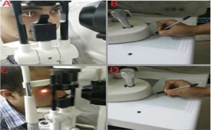
Figure 1: Slit lamp Exophthalmometry. The slit lamp is first focused on the cornea (A), and the position of the microscope is marked on the graph paper (B). Then the slit lamp is focused on the lateral orbital rim (C) and a second mark is drawn on the graph paper (D).
The Exophthalmometry value measured from both Hertel and Slit lamp were then analyzed using SPSS 23. The statistical analysis between two instruments were done using Z-test and the correlation was found using Intra class correlation.
In our study, a total of 200 subjects were evaluated. Out of which 94 were females and 106 were males. The mean age of the subject was 37.57 year with the standard deviation of ± 18.85.
|
Eye |
Technique |
Mean (mm) |
Standard deviation (±) |
|
OD
|
Hertel |
17.81 |
0.948 |
|
Slit lamp |
17.82 |
1.066 |
|
|
OS |
Hertel |
17.94 |
0.935 |
|
Slit lamp |
17.80 |
0.956 |
Table 1a: Relationship between level of awareness regarding areas of dentistry where nanotechnology has been utilized and demographic characteristics of the respondents.
*Fischer’s Exact.
|
Eye |
Technique |
Z test |
p value |
Intraclass correlation (r) |
Coefficient of correlation (%) |
Reliability (r’) (%) |
|
OD |
Hertel |
0.099 |
> 0.01 |
0.828 |
82.8 |
90* |
|
|
Slit lamp |
|
|
|
|
|
|
OS |
Hertel |
1.48 |
> 0.01 |
0.807 |
80.7 |
89* |
|
|
Slit lamp |
|
|
|
|
|
Table 1b: Comparison of Hertel and slit lamp exophthalmometry.
*: Reliable.
|
Eye Technique |
Mean (mm) |
SD |
Z-test |
p value |
|
OD Hertel |
17.81 |
0.948 |
-1.38 |
> 0.01 |
|
OS |
17.94 |
0.935 |
|
|
|
OD Slit lamp |
17.82 |
1.066 |
0.197 |
> 0.01 |
|
OS |
17.80 |
0.956 |
|
|
Table 2: Comparison of exophthalmometry value of OD and OS using Hertel and slitlamp exophthalmometer.
|
Eye |
Technique |
Gender |
Mean(mm) |
SD |
Z test |
p value |
|
OD |
Hertel |
M |
18.08 |
0.863 |
8.0210 |
<0.01 |
|
|
|
F |
17.05 |
0.948 |
|
|
|
OD |
Slit lamp |
M |
18.12 |
1.039 |
4.525 |
<0.01 |
|
|
|
F |
17.47 |
0.991 |
|
|
|
OS |
Hertel |
M |
18.17 |
0.856 |
3.879 |
<0.01 |
|
|
|
F |
17.67 |
0.955 |
|
|
|
OS |
Slit lamp |
M |
17.97 |
0.899 |
2.68 |
<0.01 |
|
|
|
F |
17.61 |
0.986 |
|
|
Table 3: Comparison of exophthalmometry value of OD and OS among male and female using Hertel and Slitlamp exophthalmometer.
The measurement of the amount of protrusion of the eye is clinically important for diagnosis, treatment and plan of management of various conditions. Thyroid Orbitopathy, Cavernous Hemangioma, Optic nerve meningioma are some of the conditions that may be associated with the axial displacement of the eye. Timely diagnosis of the conditions can improve the prognosis of the cases. Variations of 1.5 to 2.0 mm could affect patient management. A variation in measurement of 2 mm is regarded as significant during serial or relative exophthalmometry [10]. So, a simple, reliable, objective technique is required. “Slit Lamp Exophthalmometry, a novel technique” talks about the decreased examiner and patient induced error. These errors were eliminated in our study as well. Better support and stability for both the examiner and the patient throughout the procedure could be the reason for the elimination of the error. Since there are no mirror system as well, instrument induced error are also eliminated in this procedure as the mirror system could always give rise to parallax error. Also, the measurement value is independent of the type or the manufacturer of Slit lamp [9]. The study “A new method for measuring exophthalmos. The slit lamp exophthalmometer” shows that with respect to Intra and Inter observer variation during the measurement, the Slit lamp Exophthalmometer provided an improved repeatability and reproducibility value as compared to the Hertel Exophthalmometer [11].
Gender factor in ocular protrusion is controversial. Migliori., et al. [12] and Dunsky., et al. [13] reported a statistically significant increase in ocular protrusion values in males. Fledelius., et al. [14] attributed that the high value for male ocular protrusion may be because of the greater body structure in contrast, other researchers did not find a significant gender difference in ocular protrusion [15,16]. In our study, there was statistically significant difference between the two genders with male exophthalmometry value being more than that of the female. The possible limitation of Slit lamp Exophthalmometry could be the attachment of the graph in the table. Some may find it as a tiresome work.
The authors wish to thank all the patients for taking part in the study and report no conflict of interest in this work done.
Citation: Diwakar Rao., et al. “Slit Lamp Exophthalmometry, An Alternate for Hertel Exophthalmometry".Acta Scientific Ophthalmology 4.7 (2021): 02-05.
Copyright: © 2021 Diwakar Rao., et al. This is an open-access article distributed under the terms of the Creative Commons Attribution License, which permits unrestricted use, distribution, and reproduction in any medium, provided the original author and source are credited.


 Open Access by
Acta Scientific is licensed under a Creative Commons Attribution 4.0 International License
Open Access by
Acta Scientific is licensed under a Creative Commons Attribution 4.0 International License
Based on a work at https://actascientific.com
ff
