Siew Yi Ching and Gim Hin Ho
Division of Gastroenterology, Department of General Medicine, Khoo Teck Puat Hospital, Singapore
*Corresponding Author: Gim Hin Ho, Division of Gastroenterology, Department of General Medicine, Khoo Teck Puat Hospital, Singapore.
Received: March 26, 2021; Published: May 17, 2021
Citation: Siew Yi Ching and Gim Hin Ho. “Beyond the Bowel: Granulomatous Renal Pseudotumor in Crohn’s Disease. Is it Possibly an Extra-Intestinal Manifestation?". Acta Scientific Gastrointestinal Disorders 4.6 (2021): 51-53.
Extra-intestinal manifestations (EIMs) occur in patients with inflammatory bowel disease (IBD). Renal EIMs are uncommon. Even more rare are granulomatous renal pseudotumors in IBD patients. We report a possible case of renal EIM presenting as a granulomatous renal mass in a patient with Crohn’s disease.
Keywords: Crohn’s Disease; Inflammatory Bowel Disease; Extra-Intestinal Manifestation; Renal Disease; Granulomatous Disease; Inflammatory Pseudotumor
Extra-intestinal manifestations (EIMs) are seen in 25 - 40% of inflammatory bowel disease (IBD) patients [1]. EIMs commonly involve the musculoskeletal and dermatological systems and are considered primary manifestations [2]. Renal EIMs are relatively uncommon and has been reported to occur in 4 to 23% of patients [3]. They are often difficult to diagnose as primary EIMs and can provide a difficult challenge to physicians managing IBD patients. Even more uncommon are granulomatous renal masses. Prompt recognition, diagnosis and treatment are important to prevent morbidity and complications associated with them. Here, we illustrate briefly, a case of a possible renal granulomatous pseudotumor EIM associated with long-standing Crohn’s disease (CD).
This is a 60-year-old Malay gentleman with complex long-standing penetrating and fistulizing CD with ocular EIMs in the form of episcleritis. He has a history of sigmoid colon perforation requiring subtotal colectomy. He developed subsequent episodes of perianal fistulae and abscesses which needed fistulotomy, Seton insertions and drainage. The resected bowel specimen histology revealed evidence of chronicity with granulomas. His colonoscopy showed skipped colitis with ulcers. He was initially started on Azathioprine and oral Mesalazine but had subsequently defaulted follow-up and treatment. The patient’s other co-morbidities include adrenal insufficiency, history of deep vein thrombosis with pulmonary embolism, stroke and chronic kidney disease from recurrent obstructive nephrolithiasis.
He was admitted for worsening renal impairment and non-contrasted computed tomography (CT) and magnetic resonance imaging (MRI) of the kidneys showed an approximately 4 cm exophytic right renal mass with adjacent lymphadenopathy (Figure 1). A biopsy showed non-caseating granulomas with adjacent areas of fibrosis, with no associated glomerulonephritis, interstitial nephritis or malignancy. Renal biopsies for acid-fast bacilli (AFB), urinary AFB and tuberculosis (TB) polymerase chain reaction (PCR) were negative. The TB Quantiferon was positive. Chest radiograph was normal. Urine cultures grew enterococcus faecalis. A serum angiotensin-converting enzyme for sarcoidal granulomas was negative. MRI anal fistula revealed several complex internal entero-cutaneous perianal fistulae. Faecal calprotectin was < 30 ug/g.
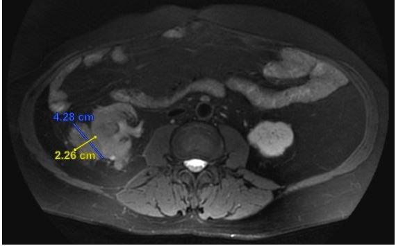
Figure 1: MRI axial view before steroids.
In light of these findings, accompanied by a history of adrenal insufficiency, corticosteroid was initiated. Intravenous antibiotics was also administered. After 2 weeks, the serum creatinine improved from 340 µmol/L (estimated Glomerular Filtration Rate (eGFR) 16 ml/min) to 235 µmol/L (eGFR 25 ml/min). A repeated non-contrasted CT kidney showed reduction in the size of the renal mass and lymphadenopathy (Figure 2 (before steroids) and figure 3 (after steroids)). Steroid was tapered with a plan to initiate azathioprine and biologics, after further consideration from the patient who had declined immunomodulators and biologics.
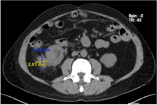
Figure 2: CT axial view before steroids.
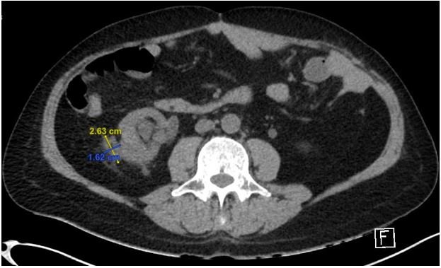
Figure 3: CT axial view after steroids.
Renal and urinary EIMs in IBD manifest primarily as nephrolithiasis, ureterocolic fistulae and ureteric obstruction [4]. Renal parenchymal disease is less common and has been reported to occur in the form of glomerulonephritis, minimal change disease, secondary amyloidosis and tubulointerstitial nephritis [4]. Granuloma formation in the kidneys is rare but they may occur in TB, sarcoidosis, Wegener’s granulomatosis and xanthogranulomatous pyelonephritis [5]. Tumor-like masses are even more uncommon and occurs almost exclusively in CD [6].
The primary differential diagnosis of a renal mass in a CD patient includes focal pyelonephritis, renal malignancy or inflammatory pseudotumor associated with CD. Following biopsy of the mass, the differentials were narrowed to focal TB pyelonephritis or Crohn’s inflammatory pseudotumor. Negative AFB and TB studies, coupled with radiological and biochemical response to corticosteroids excluded the former. While patient’s TB Quantiferon was positive, it was more consistent with a history of latent TB, rather than active TB. Wegener’s granulomatosis can be associated with CD and can rarely present with a renal inflammatory pseudotumour. However, it can be differentiated from direct renal manifestation of CD by necrotizing granulomas and vasculitis seen histologically [7], which were absent in our case. In addition, although urinary cultures grew enterococcus, biopsy excluded an active bacterial abscess. Therefore, with the evidence, this was likely a case of inflammatory granulomatous pseudotumor associated with CD.
Can this finding be considered a true renal EIM or merely an associated phenomenon? EIM is defined by an inflammatory pathology in an IBD patient that is located outside the intestine and for which the pathogenesis is either dependent on extension or translocation of immune responses from the intestine, or is an independent inflammatory event perpetuated by IBD or that shares a common environmental or genetic predisposition with IBD [8]. The exact pathogenesis of renal EIMs is unknown, but is postulated to be similar to other EIMs. They are hypothesized to be due to an extension of immune response from the intestine from ectopic expression of gut-specific chemokines and adhesion molecules, T-cell trafficking driven by non-specific adhesion molecules, microbial antigen translocation and cross-reactivity or circulating antibodies [8]. Alternative non-immune mechanisms for pathogenesis include altered hematopoiesis or dysbiosis of gut microbiota [8]. In our case, it is therefore likely that the presence of a granulomatous inflammatory renal pseudotumor is a primary EIM of CD.
Inflammatory granulomatous renal pseudotumour is a rare but important differential diagnosis of a renal granulomatous mass in patients with CD and is likely part of the spectrum of renal EIM. The morbidity associated with them is significant and there is often a small window of injury reversibility. Therefore, a high index of clinical suspicion is needed for early recognition, diagnosis and treatment to minimize short and long-term complications. This task is made more difficult by the subtle clinical presentation and that it often mimics other conditions, making a precise diagnosis difficult.
Inflammatory granulomatous renal pseudotumor is an uncommon finding in CD and possibly represents a renal EIMs of IBD. They are associated with significant morbidity. Close vigilance and early recognition remain crucial to identifying this uncommon renal EIM of CD.
Copyright: © Siew Yi Ching and Gim Hin Ho. This is an open-access article distributed under the terms of the Creative Commons Attribution License, which permits unrestricted use, distribution, and reproduction in any medium, provided the original author and source are credited.


 Open Access by
Acta Scientific is licensed under a Creative Commons Attribution 4.0 International License
Open Access by
Acta Scientific is licensed under a Creative Commons Attribution 4.0 International License
Based on a work at https://actascientific.com
ff